Patients often wonder which examination to choose — X-ray, computed tomography, or MRI. Despite having a similar purpose, these methods differ fundamentally in technology, informativeness, and indications.
X-ray: a quick basic method
X-ray remains one of the most common methods of primary diagnosis. The method is based on the use of X-ray radiation and allows for a two-dimensional image of the examined area.
X-rays are most often used to detect fractures, assess the condition of the lungs in cases of suspected pneumonia or tuberculosis, as well as to identify gross changes in organs.
Its advantages include speed of execution (a few minutes), availability, and relatively low cost. However, the method has limitations: it visualizes soft tissues poorly and does not provide a volumetric picture. Additionally, it uses ionizing radiation.
X-rays are usually prescribed for injuries, complaints of cough or shortness of breath, as well as for monitoring the condition after injuries.
Computed Tomography: detailed 3D visualization
Computed Tomography (CT) combines X-ray radiation with computer data processing, forming layered three-dimensional images.
The method allows for a more accurate assessment of the condition of bones, joints, lungs, and internal organs. It is widely used for diagnosing internal bleeding, tumors, and complex fractures.
CT is characterized by high detail and speed (on average 5–10 minutes), but it is associated with a higher radiation dose compared to a regular X-ray. In some cases, the introduction of a contrast agent is required, which has contraindications.
Examinations are often prescribed for injuries, traffic accidents, suspected strokes, as well as for clarifying oncological processes.
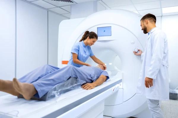
Magnetic Resonance Imaging: focus on soft tissues
MRI works without X-ray radiation — it uses a magnetic field and radio waves. This makes the method safe in terms of radiation exposure.
It is particularly effective for studying the brain, spinal cord, intervertebral discs, ligaments, tendons, and other soft tissues. Due to the high contrast of images, MRI is considered one of the most informative methods in neurology and orthopedics.
Among the disadvantages are the duration of the procedure (up to an hour), high noise levels, and limitations for patients with metal implants or claustrophobia.
MRI is prescribed for headaches, suspected neurological diseases, back pain, ligament injuries, and for diagnosing brain tumors.
Important considerations
The choice of diagnostic method depends on the clinical task and should be determined by a doctor. A more complex and expensive examination does not always mean a more accurate result in a specific situation.
In some cases, a standard X-ray is sufficient, while MRI is not a universal solution for all types of diagnosis.
X-ray, CT, and MRI are complementary methods, each of which is used depending on the suspected diagnosis. The optimal examination strategy is determined by a specialist, taking into account symptoms, medical history, and clinical picture.