This is likened to a dance or a duel, the scientists compare.
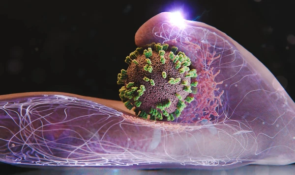
Scientists from Switzerland and Japan have created a new type of "super microscope" that combines two powerful visualization tools: atomic force microscopy (AFM) and fluorescence microscopy. They developed a new method for studying the micro-world called dual confocal AFM microscopy with visualization (ViViD-AFM). Using these technologies, scientists have for the first time in history been able to observe in real time and in high quality how the influenza virus interacts with a living cell.
The new method is advantageous as it allows for the observation of viruses without staining, enabling the study of their activities in their natural state. Almost immediately after the observations began, it became clear that science had been mistaken in many aspects of how viruses interact with cells. The general mechanism of infection has been confirmed, but its details are surprising.
For example, it is very difficult for the virus to attach to the surface of the cell — it needs large protrusions made of actin protein, which exist during the cell formation stage but then diminish. The cell, in turn, actively resists the unwelcome guest — its membrane constantly changes shape, trying to repel or block the virus. This is likened to a dance or a duel, the scientists compare, and both participants in the process behave very actively.
With ViViD-AFM, it will be possible to study how different types of viruses penetrate living cells. As well as how antiviral drugs and various vaccines do this. According to the authors of the super microscope, many new important discoveries in microbiology will be made with its help in the near future.