An international group of researchers has developed the largest reference model of the human brain to date based on data from over 54,000 individuals. The new tool allows tracking how neural connections develop and age, as well as helps identify deviations associated with cognitive impairments and neurological diseases.
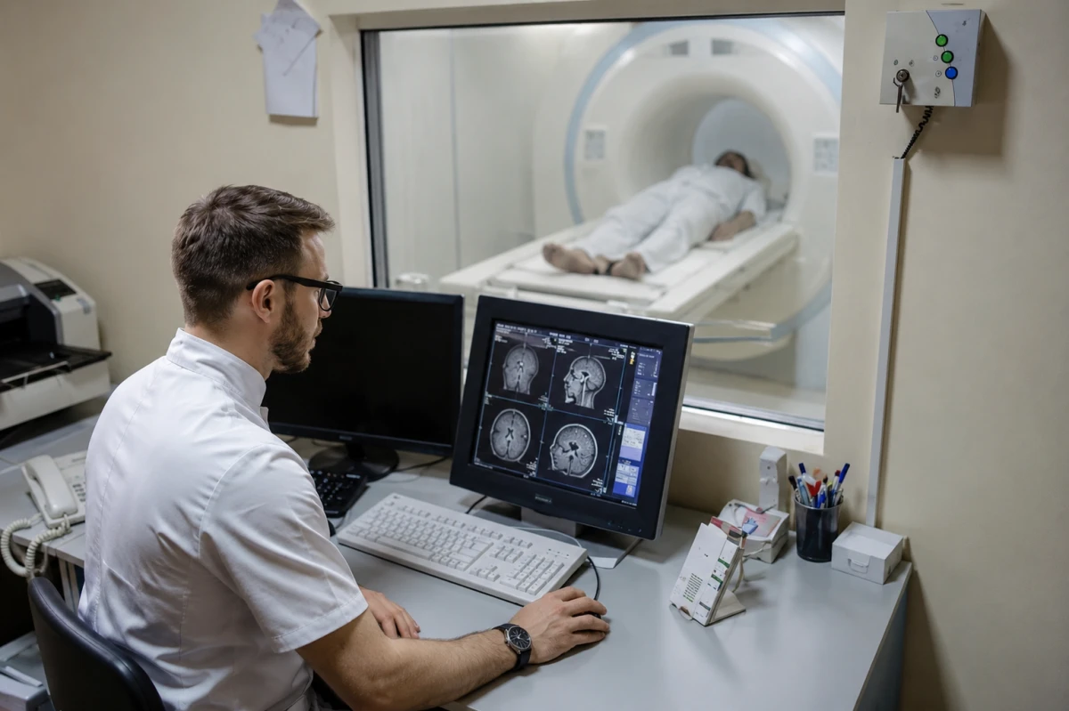
Researchers from the Mark and Mary Stevens Institute for Neuroimaging and Informatics at the Keck School of Medicine of the University of Southern California have created a large-scale reference model of the human brain. It is based on diffusion magnetic resonance imaging data from over 54,500 individuals, collected from 19 international databases.
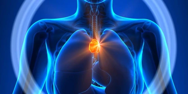
The results of the study were published in the journal Nature Communications. The authors of the research refer to their development as a kind of "atlas of development" of the brain's white matter — a complex network of nerve fibers that provides communication between different parts of the brain.
The scientists focused on four key indicators of white matter microstructure in 21 brain regions. Using modern modeling techniques, they constructed age-related curves of changes and percentile ranges, similar to growth and development tables used in pediatrics.
The resulting model allows for the assessment of the state of neural connections in relation to age norms at different stages of life — from childhood to advanced age.
One of the important conclusions of the study was the confirmation of the "last in, first out" theory. According to this concept, those areas of white matter that continue to develop the longest and reach maturity later than others are the most vulnerable to age-related degradation.
According to the researchers, this helps to better understand the fundamental mechanisms of brain aging and the reasons for age-related declines in cognitive functions.
The practical value of the new model was tested on patients with mild cognitive impairment, dementia, and 22q11.2 deletion syndrome — a genetic disorder associated with an increased risk of mental and neurological disorders.
In all cases, the system successfully identified deviations in the structure of neural connections compared to age norms. Moreover, the researchers found that even patients with the same diagnosis can have completely different patterns of brain changes.
This observation underscores the importance of a personalized approach to the diagnosis and treatment of neurological diseases.
The authors of the study believe that the created model could become an important tool for both scientific research and clinical practice. It will allow for more accurate assessments of brain development and aging processes, as well as earlier detection of signs of diseases related to neural network dysfunction.
The reference model of the brain created by the scientists opens new opportunities for studying aging processes and neurodegenerative diseases. In the future, such an "atlas" will help doctors more accurately determine individual characteristics of brain conditions, identify pathological changes at early stages, and select more effective treatment methods for each patient.