
The atlas, with a volume of about 13 terabytes, is available for free upon request.
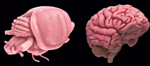
Allan Johnson from Duke University (USA) and colleagues presented a three-dimensional stereotaxic atlas of the mouse brain, covering anatomical structures and cells. To create it, the brains and skulls of five mice were visualized in three ways.
First, the brain within the skull was three-dimensionally visualized using diffusion tensor imaging with a resolution of 15 micrometers (2.4 million times higher than clinical tomographs), which allows for the examination of the cytoarchitecture of brain structures. Then, reference points on the skull were marked using micro-computed tomography. After that, the brain was extracted and images of its slices were taken using plane illumination microscopy to obtain cell maps. The results of the work were published in the journal Science Advances.
Data from all methods of visualizing the brains and skulls of the five animals were averaged, geometric distortions were corrected, and integrated into a three-dimensional atlas, with all structures labeled. It is intended to facilitate structural and functional neurobiological research conducted on the mouse brain, as well as to provide visual material for student training. The DMBA atlas, with a volume of about 13 terabytes, is available for free upon request.
Earlier, Tomas Novakowski from the University of California, San Francisco, and colleagues presented the first results of applying these approaches to studying the spatial and temporal development of brain cells — neurons and glia — and the connections between them in mice, macaques, and humans. Tracking the developmental trajectories of more than 770,000 excitatory glutamatergic, inhibitory GABAergic, and glial cells in the visual cortex of mice showed that the diversity of these cells sharply increases in the postpartum period. Large-scale mapping of 10.3 million cells in the mouse cortex in situ using the BARseq method demonstrated that their region-specific organization is influenced by changes in incoming sensory information during development.


Leave a comment